Anatomy and Physiology of Human Eye
Last Updated :
12 Jan, 2024
The human eye’s anatomy and physiology refer to its design, components, and function. The eye is a vital sense organ and is one of the five major sense organs in the human body, including the ear, nose, tongue, and skin. These organs aid in developing, learning, and adapting to our surroundings to ensure survival. The human eye can be called the living camera that transfers information about our surrounding to the brain so our brain can process this information and allow us to see the surrounding
About the Eye
The eyes are responsible for providing us with a sense of vision, which is essential for proper navigation. They are highly beneficial and unique in the human body as they can move their lens, allowing us to look around without moving our necks. The eyes work with the optic nerves to develop an image of the object we see. When light enters the eye, it forms an image of the object, which we can see and interpret.
Structure of Eye
The eye is predominantly round in shape, however, its structure is mostly covered by the Eye Leaves/lids which give it a convex appearance. It’s important to note that the Eye Leaves are not part of the eye itself. The anatomy of the eye is made up of several components working together. In addition to these components, there is a secondary structure that helps the eye function smoothly.
Human Eye
Primary Components of Eye
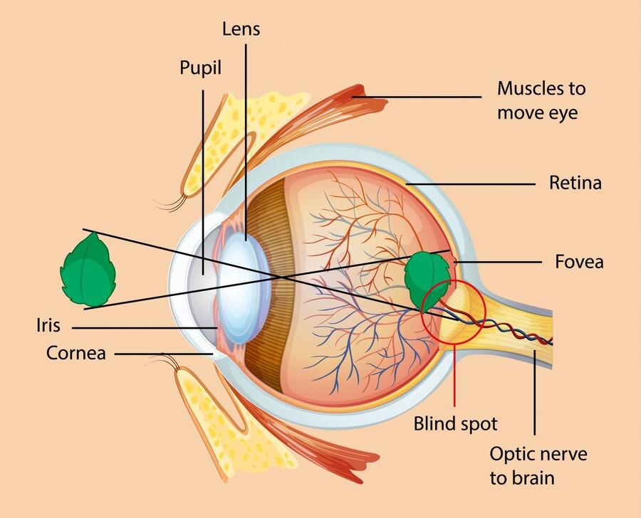
The eye has several primary components, including the cornea, sclera, aqueous humor, iris, pupil, lens, ciliary muscles, vitreous humor, and retina.
- The cornea is a circular, transparent structure located at the front of the eye, while the sclera is the white portion that covers most of the eye visible from the outside.
- The name Aqueous humor suggests ????queous” means water due to the watery nature of this area. It is a jelly-like substance that contains mostly water i.e. about 99% and about 1% vitamins and proteins. It is found between the cornea and the lens.
- The iris is a circular region in the middle of the eye made up of fiber-like structures, containing pigments (Black, Blue, Brown, etc.) that determine its color. These pigments are inherited from the parents of any individual.
- The pupil is a circular hollow in the center of the iris.
- The lens is an olive-shaped structure located behind the pupil, enclosed in a transparent capsule. It is the main component of the eye that lacks any blood vessels.
- The ciliary muscles hold the lens in its position.
- The name Vitreous humor suggests “Vitreous” means glass structure as it is transparent like glass. It is a transparent, jelly-like substance inside the eyeball that is not soft like jelly.
- The retina is the receipting component of the eye which has the only blood vessels of the eye and is connected to the optic nerve. It consists of around 130 million cells that are of two different types of cells: rod cells and cone cells.
- The cone cells work as light-sensing cells. There are nearly 5 million Cone Cells present in the eye. There are three types of cone cells; the Red Light Sensing Cone Cells, the Green Light Sensing Cone Cells & the Blue Light Sensing Cone Cells. Among them, Red light-sensing cells are present in most of the number & Blue light-sensing cells are present least in number. All other colors can be accepted by the combination of those cells.
- Rod Cells: These are also the light-sensing cells present in Retina. There are 125 million Rod Cells present in Retina. These cells can only able to accept low bright light and no bright light at all.
Secondary Components of Eye
The eye has several secondary components, including the optic nerves, and layers of the eye.
- Optic Nerves: This is the main component of the secondary eye components. This is very important for visualizing anything. Optic nerves merged with the Retina in the eyes. Nearly one million nerve fibers merged with the Retina. Optic Nerves are part of the Central Nervous System (CNS). They carry the messages from the eye to the brain.
- Layers of Eye: There are mainly three layers present in the eye. The different components of the eye merged & define such layers.
- Fibrous Tunic: It is the outer layer of the eye. It is made with tissues. And it covered the outer layer of the eyes. The outer layer of the eye is also made of Sclera & Cornea of the eye.
- Vascular Tunic: It is the middle layer of the eye. Though it is not surrounded by the eye. But it divides the eye into Interior & Posterior portions. It is also made with tissues & fibers. The Ciliary Body & Iris are two components that developed the Vascular Tunic.
- Inner Layer: The inner layer of the eye means the Retina of the eye. There are no tissues present in this area.
Functions of Eye Components
The functions of the eye components are:
- Cornea: The cornea helps to bend the light rays. As it is convex in shape, it will turn the outer light source & make convergent beams of light. It helps to pass through the Pupil of the eyes.
- Sclera: It helps to provide shape to the eyeball. It also helps to protect the eyeballs from any external threats. Also, it has the muscle Conjunctiva, which helps to move the eyeball up & down.
- Aqueous Humour: It helps to maintain the shape of the Cornea. As Cornea is convex, it provides support to the Cornea. So, the Cornea will never get misshaped. Also, it helps to reduce friction while moving the Lens of the eye.
- Iris: The light beams enter via Cornea. But the complete light beams are not necessary to get inside the eye. Iris absorbs the extra light beams & reflects them. Depending upon the pigments available in the Iris, we can see some colored Iris for individual humans. Also, Iris helps to maintain the size of the Pupil. Depending upon the environment the Pupil size gets changed.
- Pupil: It has only a single task to enter the convergent light beams inside of the eyeball. With the help of Iris, Pupil can change its size. Like in darkness, when there is a need to get more light beams inside of the eyeball, Iris helps to get stretched the Pupil. So by the Pupil, more lights enter the eyeball. In more bright light, Iris contracts the Pupil. So that minimum light beams can enter the eyeball. Otherwise, more bright light can harm the inside of the eyeball. It can able to damage the cells also.
- Lens: This helps to make a clear picture of the Retina. The lens works the same as the camera lens. As it is convex it can make convergent light beams. It helps to focus all the convergent light beams to a particular point of the Retina. So a clear picture can be available at Retina.
- Ciliary Muscles: These muscles hold the lens in its place. Also, depending upon the light beams can change the structure of the lens. If the incoming light beams are convergent, they will elongate the lens. So that, Lens can make a convergent light beam easily. If the incoming lights are divergent, the ciliary muscles compress the Lens. So it can easily focus the convergent light on Retina.
- Vitreous Humor: It is the fluid-like structure present on the inside of the eyeball. It helps to support the shape of the eyeball from the inside. Without it, the shape of the eyes makes gets misshaped due to injury.
- Retina: This helps to collect the light beams from the outside. Then, it will make the picture of the image from where the light is coming. However, the picture is dependent upon the environment of the light-receipting cells. After making the image of the object, it provides the details to the Optic Nerve.
- Leaves of the Eye: It helps to protect the eyeball from any injuries. Also, it helps to provide the shape of the eyeball.
- Optic Nerves: These nerves collect information from the retina. Then it decodes the information into the message or commands which will be transferred to the optic lobe of the brain. There the messages are interpreted by the brain & the picture of the object can be visualized by the human.
Mechanism of Eye
When light beams from outer objects enter the eye, they first land on the cornea. The cornea makes the beams converge and focuses them on the pupil. The light then travels through the aqueous humor to reach the pupil. Some of the light gets absorbed by the iris, but the rest enters the eyeball. The lens then tries to make a convergent light beam, which focuses on a specific point on the retina depending on the nature of the light beam. After being convergent by the lens, the light beam travels through the vitreous humor and lands on a special region of the retina that can make a clear picture. The picture developed on the retina is reversed from the actual object. The image is then converted to a message and received by the optic nerves, which transfer the messages to the optic lobe of the brain. The brain reads the message and develops the actual image of the object. The eye works similarly to a camera, where the iris functions as the aperture, and the retina works like the film of the camera.
What is Eye Movement?
Inside our eyes, there is only one type of muscle present i.e. the ciliary muscles. However, there are some muscles present outside of the Sclera that help to rotate the eyeball. These muscles are seven in number; levator palpebrae superioris, superior rectus, inferior rectus, medial rectus, lateral rectus, inferior oblique, and superior oblique. The cranial nerves connect the brain with the eye that sends the signal for the movement of the eyeball which leads to the rotation of the eyeball up to some degree of position. Based on the action & muscle interaction the eye movement names are generated.
- Saccades: This movement is used to see a rapid change in the position of an object. This movement involves when we don’t have the option to move the head & neck. At that point of time of gazing at a certain object which moves very fast, this Eye Movement helps us to look at that.
- Vergence: This is a special kind of Eye movement. This type of movement can visualize when we need to focus on a certain object. It helps to make a clear image in the Retina. The eye moves in such a way that light beams from that object land on the focus area.
- Smooth Pursuit: This is a very soft kind of movement in the Eye. This is a very smooth movement rather than other movements in the Eye. When we have to continuously gaze at an object which is moving in a small amount, then this movement helps us. In this movement, Eyeball rotates in a very small amount. Hence, there is no extra work that has to be done by the Eye muscles.
- Vestibulocular: This is another type of Eye movement. In some fields, where the heads move drastically. But the eye needs to focus on any object to see it. Then this type of movement can be observed. This type of movement can observe in a crowded place. These individuals need to observe certain interesting things. But due to the huge crowd, they can’t properly see them. As a result, they move their head at two different sides of their body. With that, the Eyes need to move to focus on the object.
Near Response of Eye
The Eye responds differently when individuals are seated in a moving vehicle. Initially, an object is at a distance but gradually moves closer. To obtain a clear image, the Eye adjusts its Lens structure through a process known as the Near Response. When a light beam approaches from a distance, it creates a divergent beam. The Eye adjusts the Lens width by contracting the Ciliary muscles to make convergent beams. However, as the object approaches, the Ciliary muscles elongate the Lens structure to focus incoming parallel beams. The Iris also contracts when the object is far from the individual to collect more divergent beams, but it expands when the object is close to prevent more incoming parallel beams. These are the actions of the Near Response of the Eye.
Importance of Eye
Our eyes are crucial sensory organs that allow us to see and navigate our surroundings. Without the ability to see, it would be difficult to locate objects or know where we are. Even animals with less advanced eyes have other sensing organs to help them navigate. It’s clear that the eye plays an important role in our lives and we cannot function without it. It’s not just a sensing organ but also a navigation tool. Those with vision problems know firsthand how important eyesight is. We use our eyes in every aspect of our lives, from typing messages to playing games. It makes our lives easier and allows us to experience the world around us.
Eye Irritation
Eye irritation is a term that defines some common problems related to the Eyes. In such cases, individuals can feel itching, pain, vision problem, etc. These cases are very common in humans. We can note some problems like:
- Dryness of the Eye: It is a problem in the lacrimal glands that maintain the wetness of our eyes. Due to the prolonged use of mobiles, and laptops, these cases can develop. Long use of electronic devices leads to dry eyes as the lacrimal glands can’t work properly.
- Itching of the Eye: Some common problems with the eye are related to foreign elements like pollen, dust, or chemicals that can lead to itching in the eye.
- Allergic Reaction: Human Eyes are susceptible to allergy. Allergies can result in redness and itching.
Eye Disorders
There are several disorders present in the eye. Some of them are very common in humans but some of them are very rare in case. Some of them don’t completely make a human blind but some of them can make visual impairment.
- Common Disorders with Eye
- Myopia: It is a disorder when individuals are capable to see an object which is situated near them but they are incapable to see objects which are far from them. Due to a problem with the Eye Lens, this type of problem may arise. This condition can be alleviated by the use of a corrective lens.
- Hyperopia: It is a disorder when objects which are situated far from individuals are seen clearly but the objects which are situated nearer to them are unclear. This condition can be alleviated by the use of a corrective lens.
- Cataract: It is a very common disorder in old-aged people. A thin layer of protein develops on the lens of the eye. Thus it makes a cloudy-like structure in front of the eye making the vision blocked. In this case, the lens of the eyes is removed and in place of that, an artificial lens is placed to clear the vision.
- Rare Disorders with Eye
- Coloboma: It is a disease in the eye where the tissues which protect the eye from injuries are missing. It is a very rare disease. In this disease, the chances to get injuries to the eye are much higher than the normal cases.
- Anophthalmia and Microphthalmia: These are inborn diseases. In the case of Anophthalmia, a child takes birth without the eyes. And in the case of Microphthalmia, the child has eyes but they are not functioning making the person blind.
- Usher Syndrome: It is a disease where a child has both hearing and vision impairment. Also, the child may have a problem with balance. This is a very rare genetic disease. Nearly, 4-17 per 100000 children get affected.
FAQs on Eye
Q: What is a Blind Spot of the human eye?
Answer:
The blind spot is the region in the eye where the retina gets connected with the Optic Nerves forming the optic disc in which there are no photoreceptors present to detect the light. Thus this area does not develop any image at the retina. That is why it is called Blind Spot.
Q: Is the human eye a camera?
Answer:
The human eyeball is the same as the camera, also known as camera-type eyes.
does
Q: How far can a human eye see?
Answer:
Humans can see up to 3 miles i.e., 5 kilometers.
Q: What is the possible reason for the high number of rod cells in the human eye?
Answer:
Human Eyes have nearly 125 million rod cell andnearly 5 million cone cells. According to evolutionary biologists in the ancient era the humans spend most of their time inside the caves due to which they mostly use the rod cell for vision to detect objects in low intensity of light inside the caves. This adaptation that requires more numbers of rod cells evolved into a permanent trait of the human eye thus resulting in the large numbers of rod cells.
Like Article
Suggest improvement
Share your thoughts in the comments
Please Login to comment...